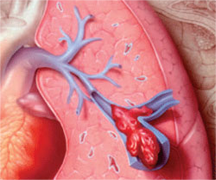
What is a pulmonary embolism ?
Pulmonary embolism is a embolism (closure) of a pulmonary artery (Arteria pulmonaria), allowing it to be fitted to the lung blood supplied by the artery which are not or only partially of oxygen. This has negative consequences for the oxygen in the lungs, to a lesser extent for the affected part of the lung itself (the oxygen supply is primarily by the bronchial arteries, because the pulmonary artery oxygen carries blood to the lungs), and - at very large terminations - for the heart. The closure consists almost always out of a blood clot, although under very special circumstances other emboli may sometimes occur (for example with air).
Pulmonary embolism causes
A pulmonary embolism is usually caused by large veins in the thigh or in the pelvis (occasionally in the heart) thrombosis occurs. The clots can break loose and then reach the lungs through the heart. Deep-vein thrombosis and pulmonary embolism are regarded as a disease.
In some diseases, such as cancer, after major operations, especially in the abdomen, and pregnancy occurs easier thrombosis causing these patients also have a greater chance of developing a pulmonary embolism. Patients with severe asthma have a nearly nine-fold increased risk of pulmonary embolism; The cause of this is not yet clear.
Large or repeated can be fatal pulmonary embolism: in Particular, the so-called "rider embolism 'whereby a large clot pulmonary arteries the shut-off of both lungs at one time, leading to a complete and immediate raises pumping action of the heart with sudden death as a result. Contrary to popular belief come from pulmonary embolism rarely veins below the level of the knee, the place Where They Usually Clearly Perceives the symptoms of thrombosis.
When puncture of a blood vessel can be accidentally sprayed air into the vessel. There is talk of a bubble That acts as embolus. Other Causes of pulmonary embolism are fat globules (or at with a break in one of the long bones), amniotic fluid constantly childbirth or decompression sickness in divers.
Pulmonary embolism symptoms
Very often little or no symptoms, especially in small embolisms. Pulmonary embolism is undoubtedly one of the most missed diagnoses. Embolism is the larger, then one can expect the following symptoms:
- Rapid and shallow breathing (92%)
- Breathlessness (dyspnoea) (84%)
- Chest pain (88%), stuck to the respiratory (74%)
- Suddenly resulting cough (53%)
- Increased heart rate (44%)
- Slight rise in body temperature (low grade fever) (43%)
- Shreds of blood in sputum (30%)
- Mortal fear
Pulmonary embolism diagnosis
The ECG shows, especially in large pulmonary embolisms, often indirect indications by the overload of the right heart half. In the blood gases often falls low pCO2 to a near-normal pO2 on (the patient is hyperventilating to maintain his O2).
Determine the radiological one made for prior use of angiography, this application is, however, largely abandoned. Nowadays is the gold standard a CT scan with contrast of the thorax (where is the embolism appears as a contrast cutout in the arteria pulmonaria or any of its branches), while also a ventilation perfusion scan qualifies as a diagnostic tool (the ventilation on the affected part of the lung will remain normal, while the perfusion decreases). The determination of D-Dimer in blood is also often carried out, however, this is a non-specific acute-phase protein and thereby, as it for example also at pneumonias is increased. This means that the positive predictive value is low. The negative predictive value of the D-Dimer is high: in the case of a negative D-Dimer determination is not treated. If the CT scan or the ventilation perfusion scan are negative, is also not treated.
Pulmonary embolism treatment
A pulmonary embolism is treated by a pulmonologist or internist at the hospital by giving anticoagulants such as coumarin derivatives and low molecular weight heparin. The treatment is aimed at preventing extension of existing clots and preventing the formation of new clots.
In case of severe, treatment-resistant hemodynamic instability, that is, a systolic blood pressure below 100 mmHg which does not respond to liquid administration by infusion, it can be carried out thrombolysis. An operation to remove the clot, or thrombectomy was found after examination less than successful thrombolysis in the acute treatment.
It seeks to potential underlying causes and the patient should use six months of anticoagulation after such an episode; Repeated pulmonary embolism even usually lifelong.
In repeated embolisms it is possible in the inferior vena cava to post a vangkorfje that captures a losschietend clot from a leg or bekkenvene can reach this heart. This technique is however only very rarely, and then only if there is a strict contraindication to treatment with coumarin derivatives or low molecular weight heparin, or if under adequate anticoagulation still occurs embolization. The reason that this operation is rarely performed is that it is a major operation with a significant operative risk, and the filter can still quickly silts and then replaced again or must be removed. In any case, the filter needs to be replaced again after 3 months.

